Anatomy and Development of the Hip
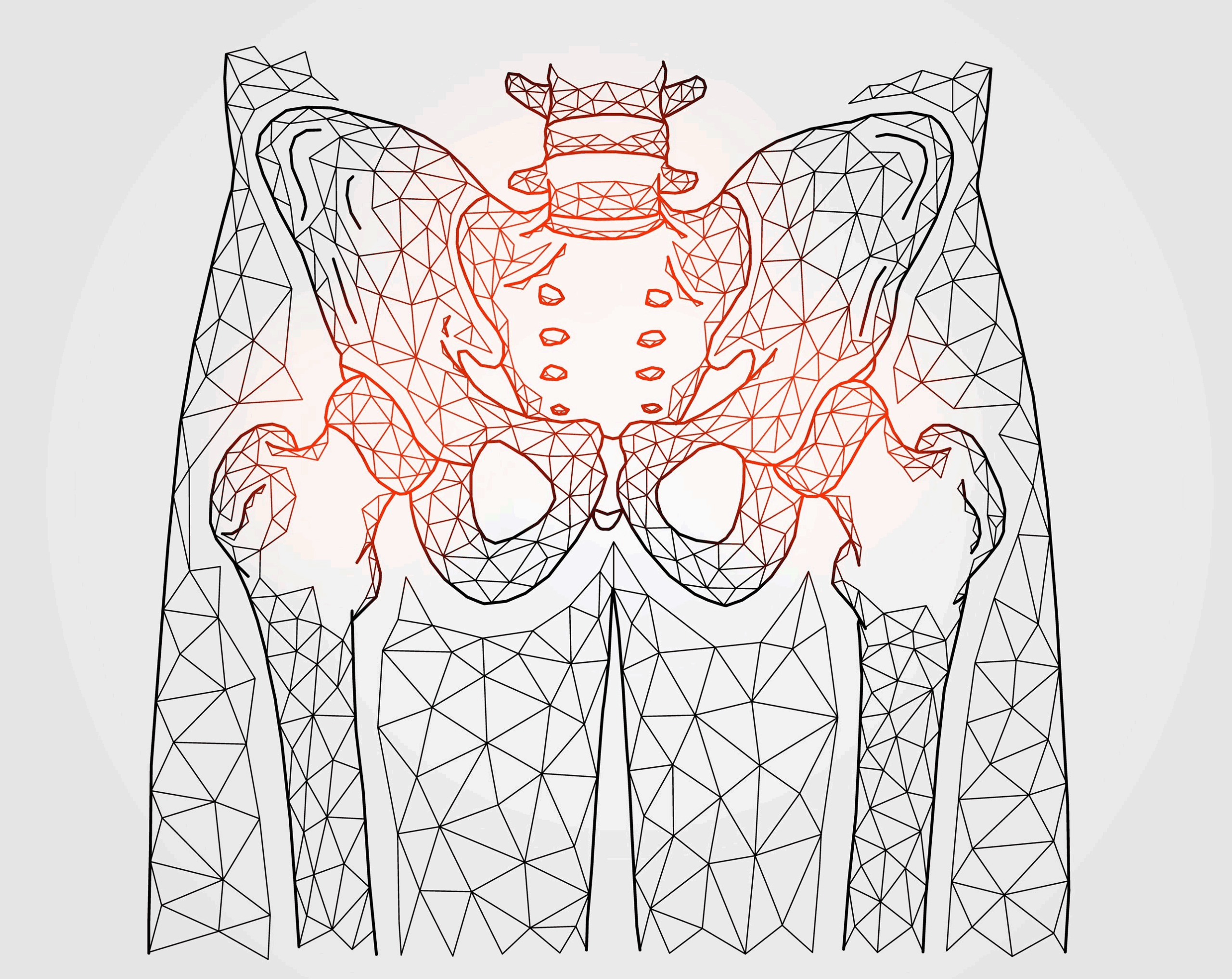
The hip joint is the connection between the head of the femur and the part of the pelvic bone known as the acetabulum. It is a ball and socket joint, allowing for a large range of motion in multiple planes. The joint is protected by articular cartilage on both the femoral and acetabular sides of the joint.
As the hip develops and matures in utero and throughout childhood, it is vulnerable to malformation and deformities, particularly to the head of the femur. The vasculature to this area is also vulnerable, as it is the only section of the body where blood flow travels from a more distal site. These deformities can result in a variety of symptoms that can be present in young childhood, or even as an adolescent.
Presentation of Hip Pathology in Children
When a patient presents with hip pain, it is important to determine the origin of the disorder, as something that presents with hip pain may be a lower back or other pelvic issue. Hip pathology itself may also present as referred knee or thigh pain, so history of the pain and clinical examination is key when suspecting a hip condition. This may be difficult in children as they may be more difficult to communicate with, especially those of a young age. It may be useful to ask the child’s parents or carers if they have noticed the child avoiding any weight bearing on that leg, or if they have a decreased level of movement. The child’s age and presentation significantly narrow down the number of diagnoses possible, and from here, imaging is very useful in confirming the specific condition.
There are three significant paediatric hip conditions that all present in different ways and at different ages. These include Developmental Dysplasia of the Hip (DDH), Slipped Capital Femoral Epiphyses (SCFE), and Perthes Disease.
Developmental Dysplasia of the Hip (DDH)
Developmental Dysplasia of the Hip, or DDH, is the name given for a spectrum of hip deformities, ranging from frank dislocation to malformation of the acetabulum leading to instability from malalignment of the joint. The hip joint is classified as Types I to IV, depending on the depth and shape of the acetabulum. Type I is a normal hip, Type II is a sublaxable, or ‘loose’, hip joint, and Types III and IV are varying degrees of dislocation.
Testing for DDH is conducted at birth through measuring the lower limbs for asymmetry, and conducting two movements, known as the Ortolani and Barlow manoeuvres. These movements test the hips’ instability and laxity, determining whether the hip is already dislocated, or is able to dislocate too easily.
The risk of a newborn developing DDH is between 0.1% and 0.5%, with an increased risk in females, those born in breech position, and those with a family history of the condition. If present at birth, it must be corrected by walking age to ensure the best possible functional outcome. Spontaneous resolution of the condition is common by eight weeks of age, though if it is still present by six months, treatment is highly recommended. The types of treatment become more extreme as the child becomes older, starting at the use of a Pavlik Harness, and ranging up to a complete hip replacement. Untreated DDH increase the risk of problems in the future, such as an abnormal gait, and increased likelihood of developing hip and knee arthritis.
Slipped Capital Femoral Epiphyses (SCFE)
Slipped Capital Femoral Epiphyses (SCFE) is the displacement of the upper femoral epiphyses, being growth plate that becomes the head of the femur, backwards off the neck of the femur. It is classified as either stable or unstable, depending on the child’s ability to bear weight on the leg in question. It has been described as looking like an ice cream scoop falling off a cone on imaging.
SCFE commonly affects males aged eleven to fourteen years of age, who are usually significantly overweight or obese. They will usually present with a limp, along with hip, knee or groin pain, though have a normal knee examination. They may also walk with the affected leg’s foot pointed outwards when progressing.
Treatment for SCFE is always required, as the vascular supply of the hip joint may become compromised, causing necrosis of the head of the femur. This treatment includes avoidance of weight bearing in the short term, and surgical fixation, or ‘pinning’. This realigns the head in the correct position, and prevents further complications. Long term follow up is required for this condition, as it is likely that the contralateral hip joint will also develop SCFE within twelve to eighteen months.
Perthes Disease
Perthes Disease is caused by the temporary obstruction of blood supply to the femoral head, leading to necrosis and death of cells in the bone. The cause of this is unknown, so treatment is very non specific and over a long period of time. It is recognised on imaging by a decrease in size and flattened edge, or an increase in density of the head of femur.
Perthes disease affects 1 in 12,000 children, and is most commonly present in males aged between four and ten years, with a peak between the ages of five and seven. Like SCFE, patients with Perthes disease will complain of hip or knee pain, and have a limp. They have a painful and limited range of rotation of the hip. Treatment for Perthes disease is rest from an aggravating activity, and surgery is only sometimes recommend. This recovery will take longer than eighteen months, and sometimes up to several years. The prognosis is dependent on the amount of the femoral head involved, with greater than 50% having a poor prognosis.