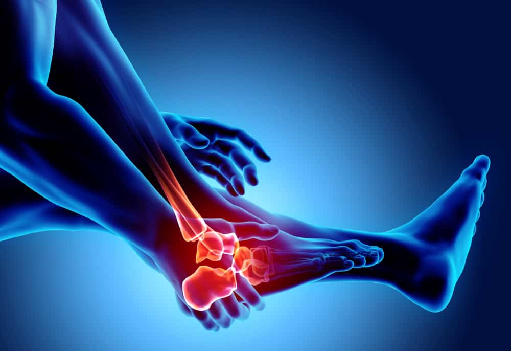
The Joint
The ankle joint is a synovial hinge joint located in the lower limb. It is formed by the bones of the leg, the tibia and fibula, and the talus of the foot. The tibia and fibula are bound together by strong ligaments and together they form a bracket shaped socket known as a mortise. The body of the talus in the foot fits snugly into the mortise, forming the ankle joint.
Movement
The ankle joint is a hinge joint. This means that it allows motion in one plane, plantar flexion and dorsiflexion. During dorsiflexion, the anterior (front) part of the talus is held in the mortise and the joint is more stable. However, during plantar flexion, the posterior (back) part of the talus is held in the mortise and the joint is less stable. You will see that you can also move your foot from side to side however, this is not due to the ankle joint but rather the subtalar joint which connects the talus and the calcaneus.
Joint Capsule
The ankle joint is a synovial joint meaning that it has a joint capsule which encases the joint architecture. The capsule contains synovial fluid which is produced by the synovial membrane. This fluid nourishes and lubricates the joint allowing it to move smoothly and painlessly.
Articular Cartilage
The joint forming surfaces of the bones that meet at the ankle joint are covered with a substance called articular cartilage. This is a slick fibre that creates friction and is present in almost all the other joints in the body. The cartilage allows the bones to slide and glide on each other without friction.
Our joints need to move regularly to remain healthy, and long periods of inactivity or immobility can increase the risk of damage to the cartilage. The articular cartilage can also be damaged by a direct heavy impact to the ankle joint and general wear and tear.
Ligaments
Ligaments are tough, flexible, fibrous tissues that act like strong ropes to connect bones to other bones, preventing too much motion and promoting stability.
| Lateral Ligaments | |
| Anterior talofibular ligament (ATFL) – The anterior talofibular ligament (ATFL) connects the tibia to the fibula. The ligament plays an important role in limiting anterior displacement of the talus and plantar flexion of the ankle. | |
| Posterior talofibular ligament (PTFL) – The posterior talofibular ligament (PTFL) also connects the tibia to the fibula. This ligament is under greater strain in full dorsiflexion of the ankle however, it is rarely injured because bony stability protects the ligaments in dorsiflexion. | |
| Calcaneofibular ligament (CFL) – The calcaneofibular ligament aids talofibular stability during dorsiflexion and restrains inversion of the calcaneus with respect to the fibula. |
| Medial Ligaments – Collectively known as the Deltoid Ligament | |
| Anterior tibiotalar ligament – The anterior tibiotalar ligament reinforces the ankle joint and controls plantar flexion and eversion. | |
| Posterior tibiotalar ligament – The posterior tibiotalar joint controls dorsiflexion. | |
| Tibionavicular ligament – The tibionavicular ligament reinforces the ankle joint. | |
| Tibiocalcaneal ligament – The tibiocalcaneal ligament also reinforces the ankle joint. |
Because the ankle joint relies on these ligaments and surrounding muscles for stability, they are easily injured. Ankle injuries occur when the ankle joint is twisted too far out of its normal position. Most ankle injuries occur either during sports or while walking on an uneven surface that forces the ankle into an unnatural position.
Muscles
Many daily activities require plantar flexion of the ankle, including walking, running, swimming, dancing, driving, and jumping. Whilst plantarflexion of the ankle seems like a simple movement, it actually requires an entire group of muscles and tendons in the leg and foot. The muscles involved are the gastrocnemius, soleus, plantaris, flexor hallucis longus, flexor digitorum longus, tibialis posterior, peroneus longus, and peroneus brevis.
Tendons
Tendons are tough bands of soft tissue that provide stability to the ankle joint. A number of tendons run through the ankle attaching muscles of the lower leg to the bones of the foot and ankle. The major tendons include the Achilles tendon which attaches the calf muscle and the calcaneus, and the flexor hallucis longus which runs along the inside of the ankle and attaches to the big toe. Most tendon injuries are the result of gradual wear and tear to the tendon from overuse or ageing. Anyone can have a tendon injury however, people who make the same motions over and over in their jobs, sports or daily activities are more likely to damage a tendon.
Bursae
A bursa is a small fluid filled sac that cushions and lubricates the ankle joint. There are three bursae in the ankle. The retrocalcaneal bursa is located in the back of the foot between the heel bone and the Achilles tendon. The subcutaneous calcaneal bursa forms at the back of the heel, and the subcutaneous bursa of the medial malleolus forms at the protrusion of the ankle where the tibia ends. Ankle bursitis occurs when bursae become inflamed. This can happen under stress from movement, from an impact injury, or even pressure on certain spots from ill-fitting shoes.